What is an OCT eye scan?
OCT screening can detect potentially serious conditions
OCT (Optical Coherence Tomography) is an advanced eye scan for people of all ages. This information will help you understand how an OCT scan can support a check-up of your eyes.

What is OCT?
OCT - Optical Coherence Tomography is an advanced eye scan for people of all ages. Similar to ultrasound, OCT uses light rather than sound waves to image the different layers that make up the structures at the front and the back of your eye. The OCT machine captures both a photograph and
a cross-sectional scan of the eye at the
same time.

What does OCT cost?
There may be an additional charge for your OCT scan, but the benefit is that the scans will give your eye care professional more information on the health of your eyes.

The scientific stuff!
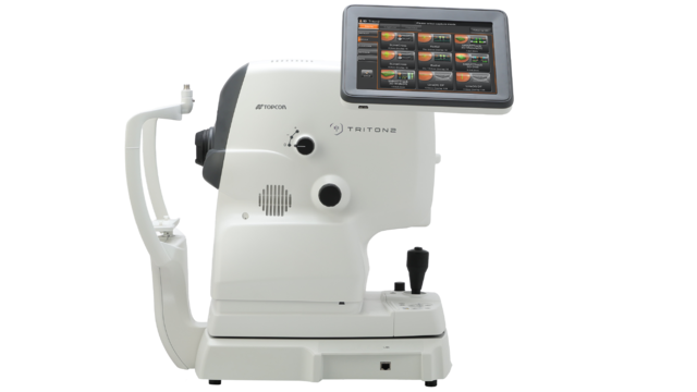
Using a Topcon state-of-the-art 3D OCT camera, your eye care professional will take both a digital photograph and a three dimensional cross sectional scan of the back of your eye in one sitting. This gives eye care professionals additional information that can assist in the diagnosis of ocular conditions. The scan is non-invasive, painless, simple and quick. Additionally, software helps to identify even the most subtle changes to the retina with every eye test you have. This gives you an invaluable ongoing record of the health and condition of your eyes.
Which conditions can OCT help identify?
Common conditions identified through regular OCT screening include:
1. AGE-RELATED MACULAR DEGENERATION
Age-related macular degeneration (AMD) is one of the leading causes of blindness globally (1). AMD causes a gradual deterioration of the macula (the central portion of your retina, enabling detailed vision). There are two types of AMD; dry and wet. Wet AMD causes a rapid reduction in vision and must be treated in hospital very rapidly. OCT can help to identify the earliest signs of AMD, determine whether it is the dry or wet form, and help monitor its progress over time.
2. DIABETES
Diabetic Retinopathy is one of the leading causes of blindness in the world (1). OCT examination helps enable early detection of diabetic retinopathy, allowing early referral and management, which can significantly improve the treatment success rate.
3. GLAUCOMA
Glaucoma is a condition that causes damage to the optic nerve – the part of the eye which connects to the brain – and causes a gradual loss in peripheral vision. Glaucoma is the second leading cause of blindness in the world. It is estimated that 57.5 million people worldwide are affected by primary open-glaucoma. (2) Because the early stages of chronic glaucoma do not cause symptoms, regular eye examinations are essential to pick up glaucoma at its earliest stage so that ongoing damage can be prevented. OCT examination can measure numerous metrics at the back of the eye and facilitate the early diagnosis of glaucoma. Furthermore, it can enable close monitoring of your eye health year on year, supporting the identification of glaucomatous changes over time.
4. VITREOUS DETACHMENTS
OCT provides invaluable information about the relationship between the vitreous and retinal surface of the eye, making it a great tool to identify and diagnose vitreomacular traction. As people get older, the vitreous jelly that takes up the space in our eyeball can change, becoming less firm and moving away from the back of the eye towards the centre. In some cases, parts do not detach and cause ‘pulling’ of the retinal surface. A vitreous detachment can be dangerous because it causes no pain, and your eye sight will seem unchanged, however, it may still be causing damage to the back of your eye
5. MACULAR HOLES
The macular is the part of the retina which is responsible for our sharp, detailed central vision. This is the vision we use when looking directly at things, when reading, sewing, or using a computer, for example. Macular holes usually form during a complicated vitreous detachment, when the vitreous pulls away from the back of the eye, causing a hole to form. Management of this condition needs to be carried out by an ophthalmologist in hospital.
1. AGE-RELATED MACULAR DEGENERATION
Age-related macular degeneration (AMD) is one of the leading causes of blindness globally (1). AMD causes a gradual deterioration of the macula (the central portion of your retina, enabling detailed vision). There are two types of AMD; dry and wet. Wet AMD causes a rapid reduction in vision and must be treated in hospital very rapidly. OCT can help to identify the earliest signs of AMD, determine whether it is the dry or wet form, and help monitor its progress over time.
2. DIABETES
Diabetic Retinopathy is one of the leading causes of blindness in the world (1). OCT examination helps enable early detection of diabetic retinopathy, allowing early referral and management, which can significantly improve the treatment success rate.
3. GLAUCOMA
Glaucoma is a condition that causes damage to the optic nerve – the part of the eye which connects to the brain – and causes a gradual loss in peripheral vision. Glaucoma is the second leading cause of blindness in the world. It is estimated that 57.5 million people worldwide are affected by primary open-glaucoma. (2) Because the early stages of chronic glaucoma do not cause symptoms, regular eye examinations are essential to pick up glaucoma at its earliest stage so that ongoing damage can be prevented. OCT examination can measure numerous metrics at the back of the eye and facilitate the early diagnosis of glaucoma. Furthermore, it can enable close monitoring of your eye health year on year, supporting the identification of glaucomatous changes over time.
4. VITREOUS DETACHMENTS
OCT provides invaluable information about the relationship between the vitreous and retinal surface of the eye, making it a great tool to identify and diagnose vitreomacular traction. As people get older, the vitreous jelly that takes up the space in our eyeball can change, becoming less firm and moving away from the back of the eye towards the centre. In some cases, parts do not detach and cause ‘pulling’ of the retinal surface. A vitreous detachment can be dangerous because it causes no pain, and your eye sight will seem unchanged, however, it may still be causing damage to the back of your eye
5. MACULAR HOLES
The macular is the part of the retina which is responsible for our sharp, detailed central vision. This is the vision we use when looking directly at things, when reading, sewing, or using a computer, for example. Macular holes usually form during a complicated vitreous detachment, when the vitreous pulls away from the back of the eye, causing a hole to form. Management of this condition needs to be carried out by an ophthalmologist in hospital.
1. World Health Organization. February 2021.
https://www.who.int/news-room/fact-sheets/detail/blindness-and-visual-impairment
2. Epidemiology of Glaucoma: The Past, Present, and Predictions for the Future
Karen Allison, Deepkumar Patel, Omobolanle Alabi Cureus. 2020 Nov; 12(11): e11686. Published online 2020 Nov 24. doi: 10.7759/cureus.11686
https://www.ncbi.nlm.nih.gov/pmc/articles/PMC7769798/
STEP 1
Book and appointment with your eye care provider
STEP 2
The eye care professional will scan your eyes using the state-of-the-art 3D OCT camera from Topcon
STEP 3
The high resolution 3D images are examined by the eye care professional using special built-in analysis tools
STEP 4
The results are presented to you
STEP 5
Any future scans can be compared with previous ones with comparative diagnosis