Dry Eye Imager
TERA
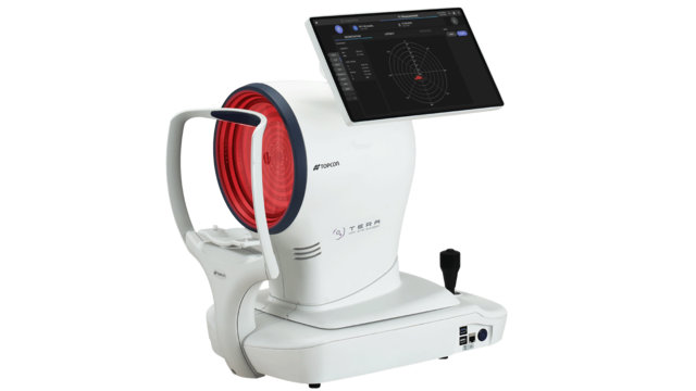
The Topcon TERA combines advanced Placido-based corneal topography with a comprehensive Dry Eye Suite – designed to support confident evaluation and management of ocular surface disease. Its automation, precision, and flexibility make it ideal for ophthalmologists, optometrists, and specialty clinics alike.
Key features
- Smart Automation: One-touch auto-alignment, focus, and capture for faster, more consistent exams.
- High-Resolution, Reflection-Free Imaging: Advanced optics and a motorised diaphragm deliver sharp, artefact-free images every time.
- Comprehensive Dry Eye Suite: Fully aligned with TFOS DEWS III, featuring non-invasive tests including NIBUT, TMH, blink analysis, lipid layer, meibography, Lissamine Green, and fluorescein imaging.
- Standardised Grading Scales: Integrated Efron, Jenvis, Guillon, and Pult scales for objective, reproducible grading.
- Corneal Topography and Screening: Detailed anterior corneal mapping with keratoconus detection and Zernike aberration analysis.
- Flexible Reporting and Review: Generate structured reports directly on the device or any connected PC using TERA Review Software.
LIPID LAYER ASSESSMENT
High-quality imaging under diffuse lighting, using an innovative, patented approach, enables non-invasive, qualitative evaluation of lipid layer flow across the corneal surface, supporting identification of abnormalities of the lipid layer as a driver of dry eye disease. Classification is supported by comparison with the internationally recognised Dr Guillon scale1.

OCULAR REDNESS ASSESSMENT
High-resolution colour imaging and motorised diaphragm enable high-quality and consistent assessment of conjunctival redness, exploiting internationally validated grading scales (Efron2 and Jenvis3) to support clear visual documentation, comparison over time and investigation of inflammation as a possible driver of dry eye.

BLEPHARITIS ASSESSMENT
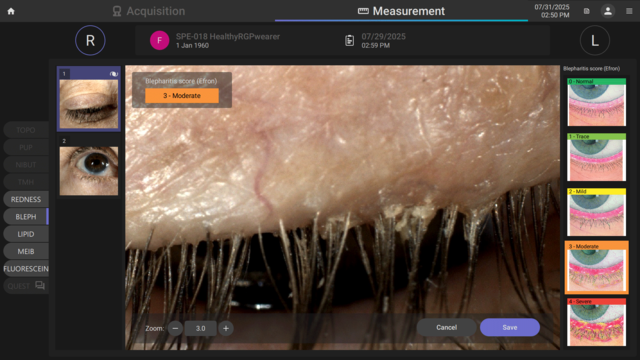
High magnification colour imaging of the eyelid margins allows clear visualisation of desquamation, inflammation, and structural changes. The images can be graded using the Efron scale2 to support consistent assessment and documentation of blepharitis inflammation severity, and investigation of lid margin abnormalities as a driver of dry eye disease.
FLUORESCEIN IMAGING AND VIDEO
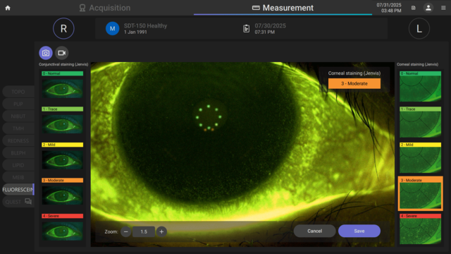
Fluorescein imaging supports the examination of ocular surface integrity and tear film stability. Staining patterns can be assessed and compared using the international Jenvis grading scale. The same imaging and video mode can also be used to evaluate and document the fit of rigid contact lenses.
DRY EYE QUESTIONNAIRES AND REPORTS
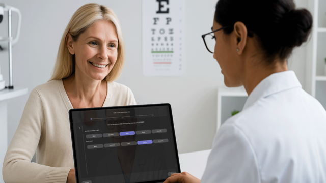
TERA includes integrated OSDI-6 (as recommended by DEWS III4), and DEQ-5 questionnaires to capture patient-reported symptoms. Combined with structured dry eye and follow-up reports, these tools support consistent assessment, reinforce treatment compliance, and help clinicians monitor changes over time.
1 Guillon, M., 1998. Use of the Tearscope Plus in the routine contact lens fitting practice. Contact Lens and Anterior Eye, 21(Suppl 1), pp.S31-S40.
2 Efron, N., 1998. Grading scales for contact lens complications. Ophthalmic and Physiological Optics, 18(2), pp.182-186.
3 Jenvis, D.R., et al., 2007. The development and validation of the Jenvis Dry Eye Questionnaire. Optician, 233(6091), pp.16-21.
4 Wolffsohn, J. S., Benítez-Del-Castillo, J., Loya-Garcia, D., Inomata, T., Iyar, G., Liang, L., Pult, H., Sabater, A. L., Starr, C. E., Vehof, J., Wang, M. T., Chen, W., Craig, J. P., Dogru, M., Quinones, V. L. P., Stapleton, F., Sullivan, D. A., Jones, L., & TFOS collaborator group (2025). TFOS DEWS III Diagnostic Methodology. American journal of ophthalmology, S0002-9394(25)00275-2. Advance online publication. https://doi.org/10.1016/j.ajo.2025.05.033
Not all products, services or offers are approved or offered in every market, and products vary from one country to another. Contact your local distributor for country-specific information.