Ultra-widefield Retinal Slit Scanner
GAIA
The GAIA brings a new era of ultra-widefield imaging, where true colour delivers a natural* view of the retina. With IR alignment and fully automated sequential bilateral capture, GAIA enables consistent imaging with minimal operator input, enhancing the patient experience while keeping clinical workflow efficient and reliable.
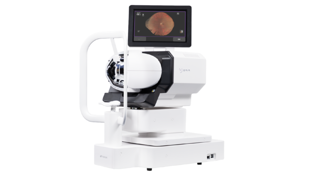
Key features
- Capture more with 209°** ultra-widefield of view
- Clinical confidence with true colour imaging
- Effortless operation, auto-alignment and capture
- Improved patient comfort
- Seamless integration for efficient interoperability
- Space-saving
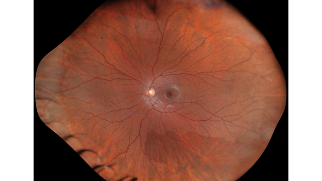
True colour
Utilising LED white light source, GAIA captures ultra-widefield retinal images in true colour1.
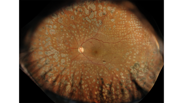
The slit-scan illumination and rolling shutter mechanism enable GAIA to capture true colour ultra-widefield fundus images through pupils as small as 2.0 mm2, supporting high-quality, efficient non-mydriatic imaging even in bright clinical environments.
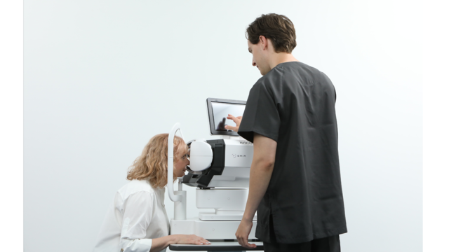
GAIA is designed to make ultra-widefield imaging simple and effortless. Real time IR imaging, automated alignment and capture, and non-contact imaging reduce the need for complex positioning and manual adjustments.
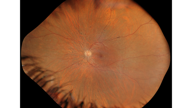
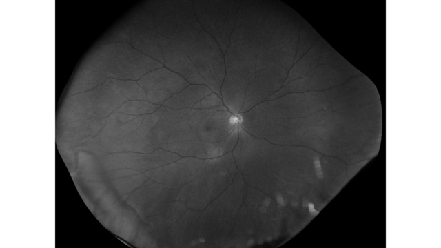
Red-free image from GAIA
Research has shown that red-free fundus imaging supports detailed assessment of retinal vessels, haemorrhages and the retinal nerve fibre layer (RNFL).
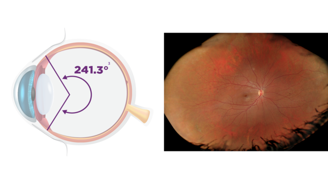
Mosaic image
IMAGEnet7 enables the generation of mosaic images.
* Refers to colour tone comparable to true colour images captured by conventional fundus cameras.
** 209° is an estimated value derived from a visual angle of 150° (GAIA), based on the visual‑angle-to‑eye‑angle relationship described in the following article. Xincheng Yao, Devrim Toslak, Taeyoon Son, Jiechao Ma. Understanding the relationship between visual-angle and eye-angle for reliable determination of the field-of-view in ultra-wide-field fundus photography. Biomedical Optics Express, 2021 Sep 30;12(10):6651-6659
1 True, full colour fundus image simultaneously captured with white light, 24-bit colour.
2 Confirmed with model eye.
3 241.3° represents an estimated eye-angle for a mosaic image, derived from a visual angle of 180° (150° central field plus two additional 15° shifts using internal fixation). The conversion is based on the relationship described in the following article. Xincheng Yao, Devrim Toslak, Taeyoon Son, Jiechao Ma. Understanding the relationship between visual-angle and eye-angle for reliable determination of the field-of-view in ultra-wide-field fundus photography. Biomedical Optics Express, 2021 Sep 30;12(10):6651-6659
Not all products, services or offers are approved or offered in every market, and products vary from one country to another. Contact your local distributor for country-specific information.