
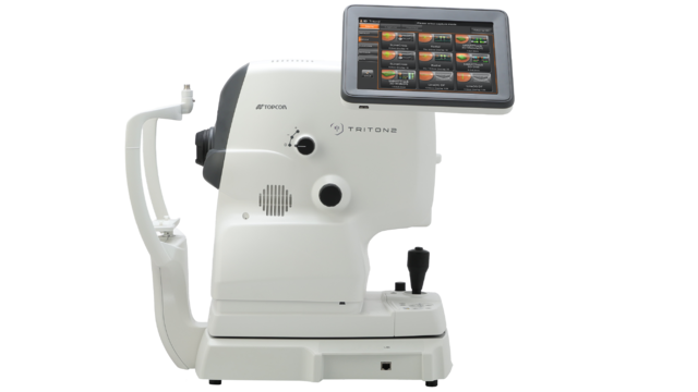
Next-Generation Multimodal Swept Source OCT
Triton2
The new DRI OCT Triton2 from Topcon Healthcare combines advanced Swept Source OCT technology with a true colour fundus camera.
Key features
- Swept-Source OCT providing high density scans and deep penetration
- Slit-scan technology to capture fundus images through small pupils (φ2.0mm or larger*)
- Wide-field OCT and OCTA, up to 21mm**
- Smart Denoise** provides higher signal-to-noise ratio on 3D OCT and OCTA**
- Flexible positioning for easier acquisition
- Powered by IMAGEnet®7
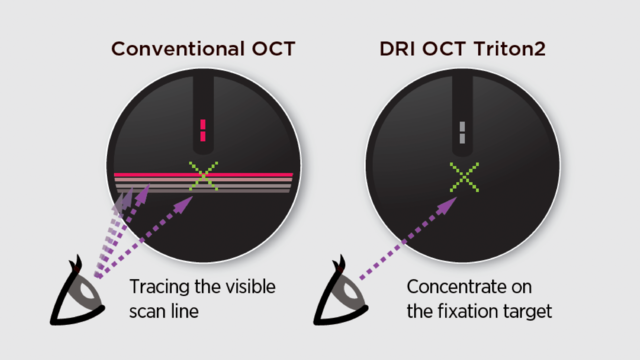
Invisible Scan Lines
The invisible 1,050nm wavelength light helps patients concentrate on the fixation target during the scan, reducing involuntary eye movement.
It supports more efficient workflow in practice by reducing the need to rescan.
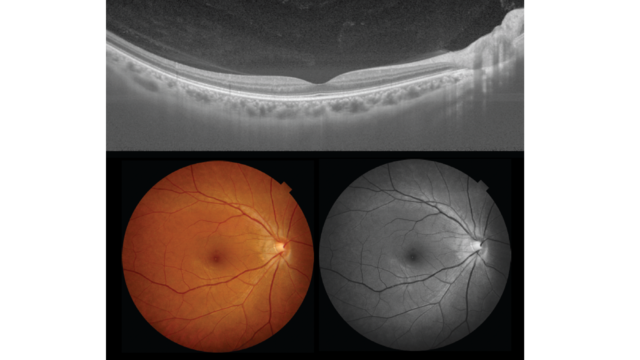
The innovative slit-scan illumination and rolling shutter mechanism in the Triton2 produces excellent quality colour fundus images with less flare and shadow.1
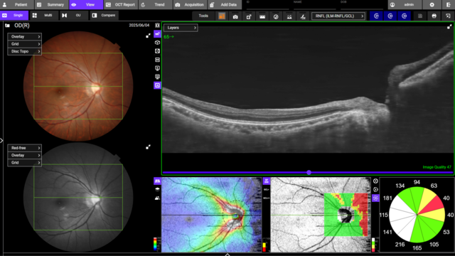
3D Wide Scan for Complete Posterior Pole Assessment
The 12x9mm 3D Wide scan captures both the optic nerve and macula in a single acquisition, delivering a thorough evaluation of the posterior pole. The Triton2 reference database extends across the entire scan area, enabling detailed thickness comparisons within the visual field, ideal for detecting patterns such as thinning of the RNFL commonly seen in glaucoma2.
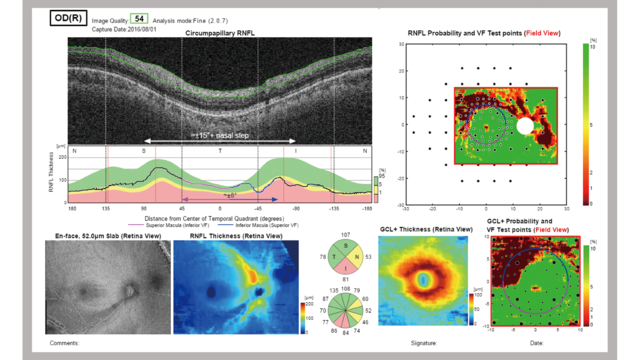
The Hood Report for Glaucoma
The Hood Report streamlines the decision-making process through the correlation of structure (GCL/RNFL) with function (overlay of visual field test locations)3. A single wide-field OCT scan with Hood report can provide compelling information for the diagnosis of early glaucoma4.
Topcon's SS OCT Angiography
The optional Topcon’s SS OCT Angio™ integrates OCT Angiography with Swept Source technology, and the long 1050nm wavelength. Powered by OCTARA™, a proprietary image processing algorithm, SS OCT Angiography enables detailed visualisation of vascular structures and the monitoring of key retinal pathologies.
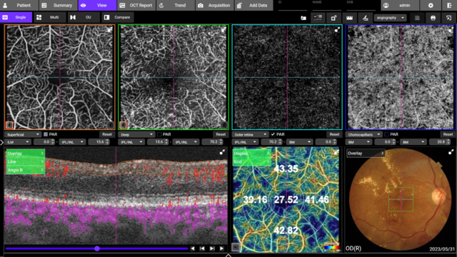
Wide-field Imaging
The optional wide-field attachment lens enables the capture of scans up to 21mm in length. Gather more clinical insights with wide-field OCT and OCTA imaging - valuable in a wide variety of conditions.

Retinal and Choroidal Thickness Maps
IMAGEnet®7 provides up to 5 retinal thickness maps, enabling quantification of retinal layers and sub-layers. The Triton2 provides clear visualisation of the choroid and generates choroidal thickness maps, supporting clinicians in gaining a clearer understanding of choroidal structural changes.
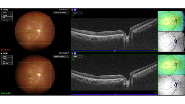
Follow-Up Function
The follow-up function enables easy retrieval and re-analysis of the same location, allowing seamless comparison of past and current data. Operators simply select previous scan data, and Triton2 automatically captures the corresponding area.
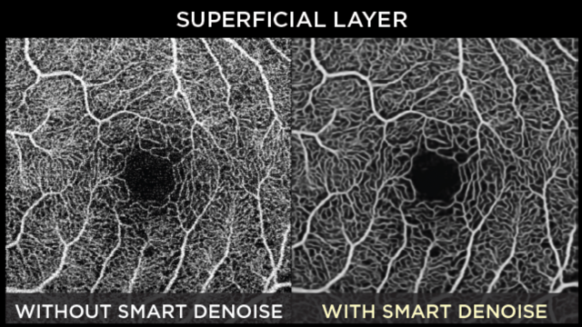
Smart Denoise**
Smart Denoise is image processing algorithm which reduces artifacts and increases contrast. High quality OCT and OCTA images with reduced noise signal are generated from every B-scan within the dense data cubes, through the use of Topcon's unique AI algorithm.
Anterior Segment Imaging
The optional anterior segment imaging capabilities allow for visualisation of the cornea, anterior chamber angle, iris and anterior sclera. The anterior segment lens attachment is combined with quantitative analysis. With the addition of the optional anterior segment features, Triton2 provides a valuable solution for comprehensive (anterior and posterior) eye care settings.
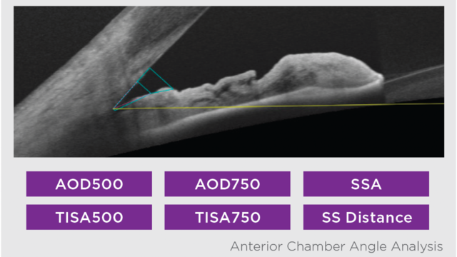
Comprehensive Assessment with Quantitative Analysis
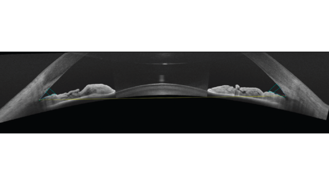
Line scan length in 16mm
Delivering a 16mm wide scan, the Triton2 captures both iridocorneal angles in a single acquisition, enabling fast and efficient evaluation. Paired with IMAGEnet7, it delivers angle measurements.
Radial scan length in 9mm
From the 9mm radial scan, the quantitative analysis provides automatic measurement of the total corneal, epithelial and stromal thickness, enabling the diagnosis and monitoring of various conditions.
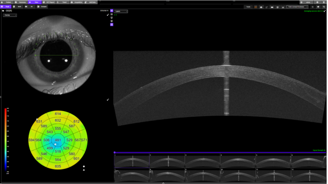
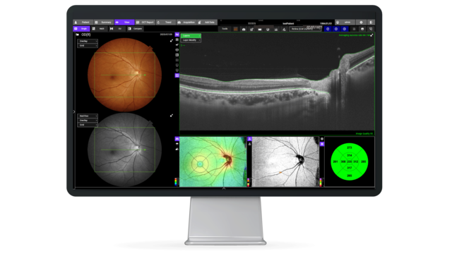
Triton2 Acquisition Control
In combination with IMAGEnet®7, the operator can view the live image on a large desktop monitor during acquisition.

* Confirmed with model eye
** Optional
1 As compared to Topcon conventional non-mydriatic retinal camera TRC-NW400
2 Comparison of glaucoma-diagnostic ability between wide-field swept-source OCT retinal nerve fiber layer maps and spectral-domain OCT Won June Lee, Ki Ho Park et al, Eye volume 32, 2018. Diagnostic Accuracy of Wide-Field Map from Swept-Source Optical Coherence Tomography for Primary Open-Angle Glaucoma in Myopic Eyes.Yong Woo Kim, Jinho Lee, Jin-Soo Kim, Ki Ho Park. AJO, 2020
3 Donald C. Hood PhD, Translational Vision Science & Technology No.6 Vol.3 2014: Evaluation of a One-Page Report to Aid in Detecting Glaucomatous Damage.
4 A Single Wide-Field OCT Protocol Can Provide Compelling Information for the Diagnosis of Early Glaucoma Donald Hood et al, 2016 Translational Vision Science & Technology
Not all products, services or offers are approved or offered in every market, and products vary from one country to another. Contact your local distributor for country-specific information.
Related Products
Ready to learn more? Contact us!
Please fill out the form below and your local Topcon Healthcare representative will be in touch soon.