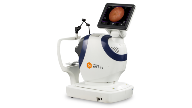
Non-Mydriatic Retinal Camera
NW500
Introducing Topcon's next-generation fundus imaging camera that offers bright and sharp images in well-lit conditions, even for eyes with small pupils.*
Key features
- Sharp, high-quality images
- Small pupils, 2mm or larger*
- Easy to use
- Robotic
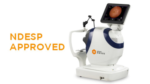
APPROVED BY THE NDESP
The NW500, has been included in the list of approved cameras for the NHS Diabetic Eye Screening Programme since March 2024. The NW500, characterized by its user-friendly one-touch capture system, enables rapid and efficient acquisition of high-quality retinal images, even in eyes with opacities and poor dilation, thus enhancing the effectiveness and workflow of busy diabetic eye screening clinics."
The DES Programme is a beneficial annual eye screening service to check for eye problems caused by diabetes, and valuable to so many patients to help detect these early changes using fundus photography.
Excellent quality fundus image
Capable of capturing fundus images through a pupil diameter as small as 2.0mm*, it provides sharp, detailed colour fundus images in dark and brightly lit rooms2.
Ease of use
Alignment, focusing, shooting, and movement between the right and left eye are operated automatically with a single touch, making the NW500 easy to operate4.


Innovative slit-scan illumination and rolling shutter mechanism in the NW500 produces excellent quality colour fundus images with less flare and shadow5.
The slit-scan mechanism helps to overcome one of the known causes of poorly graded images with its ability to effectively image through small pupils. This innovative technology also helps the NW500 capture sharp, high-quality fundus images, regardless of miosis and the lighting conditions, unlike conventional fundus cameras.

Panoramic wide-field photography
In peripheral photography mode, it is possible to create a wide panorama image with an approximately 90-degree angle of view (approx. 132.3 degrees at the spherical centre of the eye3).


*φ2.0 mm or larger, tested on model eye.
2 A room with a brightness of 623 lux or less.
3 Xincheng Yao, Devrim Toslak, Taeyoon Son, Jiechao Ma. Understanding the relationship between visual-angle and eye-angle for reliable determination of the field-of-view in ultra-wide field fundus
photography. Biomedical Optics Express, 2021 Sep 30;12(10):6651-6659.
4 If the auto-shoot setting is turned on.
5 As compared to Topcon non-mydriatic retinal camera TRC-NW400.
6 Multiple connections with Direct DICOM, Ez Capture, IMAGEnet® 6, Shared folder and Direct Storage (USB/LAN).
Not all products, services or offers are approved or offered in every market, and products vary from one country to another. Contact your local distributor for country-specific information.